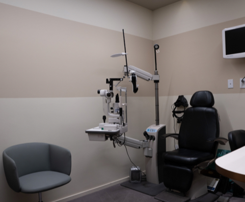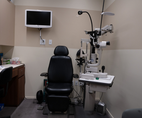
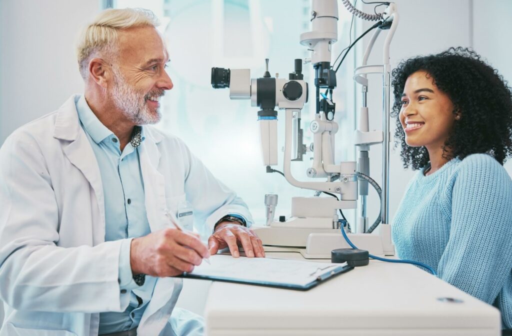
Here at Orchard Park Optometry, we always want to improve how we can serve our community. That’s why we’re always trying to incorporate new technology into our comprehensive eye exams; we can be more in-depth during the exam to learn more about what’s going on inside your eyes.
When you come in for an eye exam with us, we’ll use the following technology:
- Optomap
- OCT
- Corneal topography
- Axial length measurement
- Digital phoropter
Eye exams are a crucial part of preserving your vision—and this technology helps us keep your eyes strong and your vision clear.
Optomap
Inside your eye, you have a small piece of light-sensitive tissue called the retina. It receives the light from your surroundings and sends it through the optic nerve. Think of it like a camera; it builds the images that let you interpret your environment.
This retina is made up of several small parts—each one as important as the next. But just like a camera, sometimes problems can develop with one or more of these parts, and this can have long-lasting effects on your vision.
This is where Optomap Ultra-widefield Retinal Imaging comes into play. This technology lets us take a high-resolution picture of your retina, letting us look for all kinds of potential problems like:
- Age-related macular degeneration
- Glaucoma
- Retinal detachment
- Cataracts
- Diabetes
- High blood pressure
This is a quick, painless, and simple procedure—it’s just like having your picture taken. Then, the image is 3D rendered, and our team will work with you to explain what’s occurring inside your eye. Optomap imaging gives us an incredibly in-depth view of your eye and helps us keep a close watch on any changes.
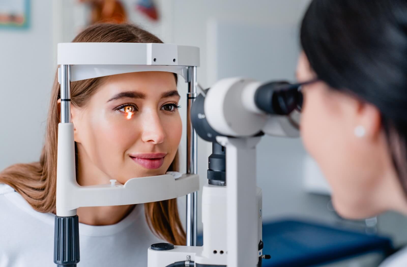
OCT
Optical Coherence Tomography, or OCT, is a quick scan that gives us a deep understanding of your retina. It’s a non-invasive scan that provides us with a hospital-quality cross-sectional rendering of the inside of your eye.
With OCT, we can meticulously examine each layer to detect early signs of conditions such as:
- Macular hole
- Epiretinal membrane
- Multiple sclerosis
- Elevated spinal fluid pressure
Using light waves, OCT captures detailed images of your retina, which our team can then analyze in real-time. This procedure helps us track any minute changes over time, ensuring we catch any potential issues as early as possible. It’s like having a magnifying glass on the microstructures of your eye, helping us keep your vision crisper and clearer for longer.
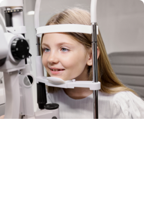
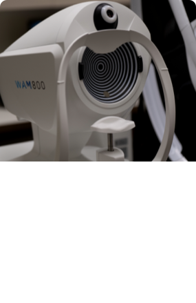
Corneal Topography
Your cornea—the clear, dome-shaped tissue at the front of your eye—is responsible for refracting light to reach the retina. To do so, it has to be properly curved, but this doesn’t mean you’re always lucky enough to have a perfectly-shaped cornea.
This is where corneal topography comes into play. At Orchard Park Optometry, we use the Topcon CA-800 Topographer to map the layout of your cornea and look for any potential problems.
Using this imaging, we can:
- Chart out any corneal defects or abnormalities
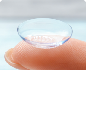
- Fit you for contact lenses like ortho-K, RGPs, and scleral lenses
- Diagnose and monitor changes in astigmatism
- Plan and monitor any refractive surgery like LASIK, PRK, or cataract surgery
- Detect keratoconus or other progressive corneal diseases
This is an invaluable and non-invasive tool that lets us gain a proper understanding of your eye’s shape—and whether or not something is wrong.
Axial Length Measurement
Think of the last time you held a magnifying glass. They don’t work perfectly at every distance; in order to properly bend light so you can see through it clearly, the lens has to be at a set distance from something. Otherwise, whatever you’re looking at is blurry and impossible to properly see.
Your eye works similarly. There’s a lens at the front that refracts light to reach the retina. This lens has to be at a certain distance from the retina, just like the magnifying glass; if it’s too close or far, you’re going to struggle to see.
This is the case with conditions like myopia (nearsightedness) and hyperopia (farsightedness). The eye grows to be too long or too short, causing blurry vision, headaches, and more. Your eye can’t refract light like it needs to.
Because of these conditions, we measure your axial length during your eye exam. This lets us have a better understanding of your specific vision troubles, and monitor any changes in your eye. By measuring your axial length, we can:
- Diagnose, monitor, and treat conditions like myopia and hyperopia
- Precisely calculate the power of intraocular lenses (IOLs) before cataract surgery
- Track changes over time to help manage conditions like glaucoma
This process doesn’t take long, and it helps us make sure we’re giving you the personalized, precise eye care that you deserve. With axial length measurement, we can keep a close watch on the growth and changes of your eye, making sure that every aspect of your vision care is finely tuned for optimal health and clarity.
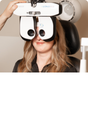
Digital Phoropter
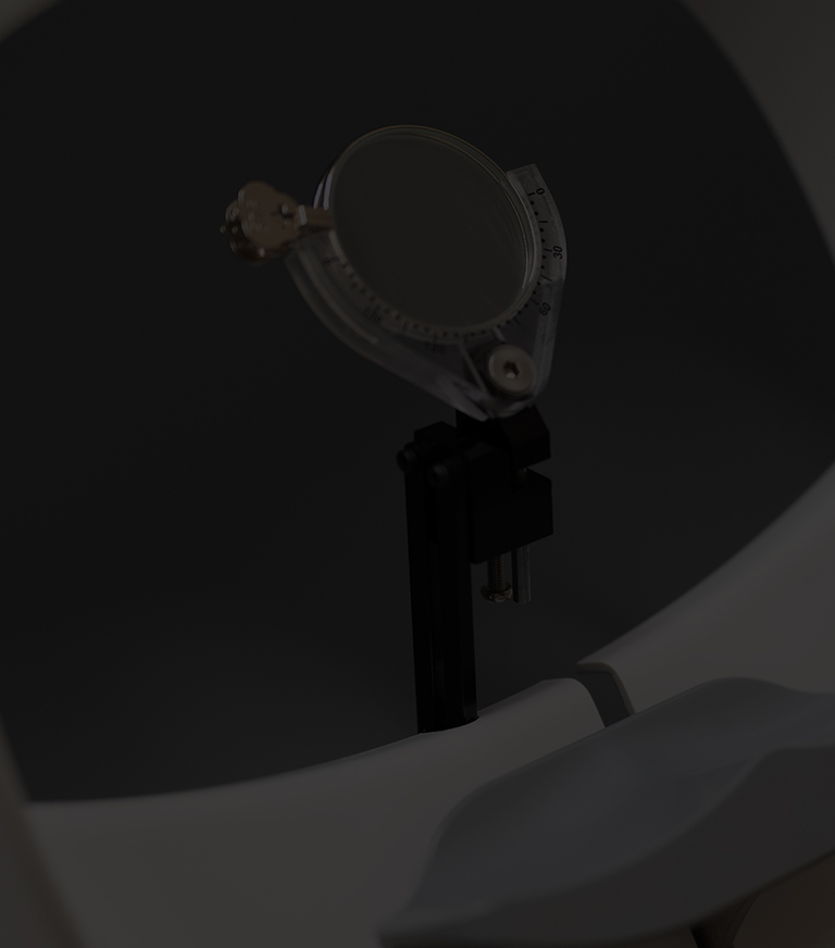
If you think back to your last eye exam, you might remember that machine that flips between several different lenses. Your optometrist will switch between lenses 2 at a time and have you compare which provides you with clearer vision. This is a phoropter—it lets us get a quick and accurate measurement of your prescription.
At Orchard Park Optometry, we take it one step further; the process is now digitalized. This helps us get an extremely precise prescription to give you the clear vision you deserve. With our digital phoropter, we can swiftly and seamlessly switch between lenses, allowing us to fine-tune your prescription with exceptional accuracy.
This reduces how long you’re in the exam room and streamlines the overall experience. Plus, the digital phoropter easily stores your prescription history, making it much easier to compare past and present results. It’s fast, efficient, and precise!
Book Your Next Eye Exam Today!
The future of eye care is exciting, and here at Orchard Park Optometry, it’s already within your sight. Using this advanced technology lets us see deeper and get a better understanding of what’s going on inside your eyes. Book your next eye exam with our team today; your eyes will thank you!